An Abnormal Lymph Node Demonstrates Which One of the Following
Looking back this phenomenon was documented occasionally as a. Left axillary lymph nodes demonstrate very subtle cortical thickening which could be related to reactive inflammation.

Ultrasonogram Characteristics A Abnormal Cervical Lymph Node Download Scientific Diagram
These data do not agree with the results of an anatomical study 8 in which the mean number of superficial and deep lymph nodes dissected at autopsy was 1360 per side range 5 -17.

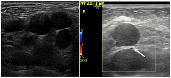
. You suspect a dentalperiodontal infection. The US examination for axillary pain demonstrated lymph node with abnormal cortical thickening of 6 mm Fig 5 B. The most common lumps or swellings are enlarged lymph nodes.
CBC is normal except for findings of mild anemia. Infected Lymph nodes tend to be firm tender enlarged and warm. This cancer occurs in the middle of the thyroid where the secretions are from.
However abnormal IMLN features such as diminished or absent hilum thickened cortex not circumscribed margins increased size or interval change warrants additional follow-up or pathologic analysis to exclude malignancy. 22 females 14 males mean age 25 years. The thoracic duct would be severed.
3-mm-thick contiguous slice matrix 512 512. The combination of bilateral hilar and paratracheal node enlargement usually allows the differentiation of sarcoidosis from lymphoma. Lymph nodes may be reactive and are not enlarged by size criteria.
The mesorectum nodes should be considered as abnormal when they are over 4 mm in short diameter. Swollen salivary glands under the jaw may be caused by infection or cancer. Swollen lymph nodes in the anterior neck usually indicate.
Lumps in the muscles of the neck are caused by injury or torticollis. No suspicious sonographic abnormalities are identified. The absence of suspicious lymph nodes must also be documented.
Nodes are not enlarged by size criteria within either axilla. Short axis diameters of pelvic and inguinal lymph nodes were measured. These can be caused by bacterial or viral infections cancer malignancy or other rare causes.
A small but not insignificant number of people develop unilateral axillary adenopathy in the days following vaccination against SARS-CoV-2 the virus that causes COVID-19. A patient presents with enlarged tender submental lymph nodes. A balanced fast field echo b-FFE sequence was used with the following parameters.
In general any vaccine could cause this sort of reactive lymph node enlargement. When abnormal lymph nodes are present on a mammogram ultrasound CT or MRI scan For some people with cancer such as breast cancer or melanoma to see if the cancer has spread sentinel lymph node biopsy or needle biopsy by a radiologist. A woman has her right breast and right axillary lymph nodes removed.
It metastasizes quickly and will show up in surrounding lymph nodes. What does a muscle knot look like. Which of the following might occur.
If present enlarged or sonographically abnormal cervical lymph nodes must be described and localized with particular attention to anatomic landmarks that can be used during FNA or neck dissection. Abnormal lymph node enlargement tends to commonly result from infection immune response cancer and less commonly due to infiltration of macrophages filled with metabolite deposits eg storage disorders. The lymph node measured 9 mm in short axis and 19 mm in long axis.
Malignant nodes have been noted to demonstrate eccentric or absent hilar vascularity multifocal aberrant vascularity peripheral perfusion focal perfusion defects or peripheral subcapsular vascularity in essence much like other cancers as they become more malignant in appearance the more abnormal the vascular flow into the lymph node becomes. Minimum lymph node involvement with deposits between 02 and 2 mm micrometastasis and 02 mm isolated tumor cells is not related to significant morphological changes in the lymph node thus limiting the usefulness of ultrasonography in such cases so the diagnosis is made by means of histology or immunohistochemistry. Reactive unilateral axillary adenopathy.
One of the signs of illness is the absence of a fatty filter inside of a lymph node which can be revealed on an ultrasound. Cisterna chyli flow increases. Examination there is an enlarged nontender supraclavicular lymph node and enlargement of the Waldeyer ring of oropharyngeal lymphoid tissue.
Clinical features of abnormal lymph node enlargement. There was no increased cortical vascularity. So on the lymphadenitis or inflammation of the lymph nodes in the clavicle can indicate the swelling of the tissues in the supraclavicular fossa visually expressed in the asymmetry of the shoulders redness and increase in the temperature of the skin at the site of the lesion pain syndrome with the movement of the shoulders and neck also felt during.
Lymph drainage would be affected in her left arm. There is no hepatosplenomegaly. A total of 730 lymph nodes were observed for a mean of 588 2009 per station and individual patient range.
Stereotactic biopsy of the left breast asymmetry revealed fibrocystic change with pseudoangiomatous stromal. A recent study examining the ability of mri to detect axillary metastases abnormal lymph nodes were defined as 1 cm matted andor exhibiting loss of fatty hilum thickened cortex andor irregular node contour in nac patients found that mri was reasonably accurate with a negative-predictive value of 78-81 and positive-predictive value of. Right lymphatic duct drainage decreases causing edema in the right arm.
A lymph node biopsy specimen shows replacement by a monomorphous population of large lymphoid cells with enlarged. This can be benign as rheumatoid arthritis and some autoimmune system disorders can cause this but it is also an early sign of cancer that would need biopsy to be certain. Swollen lymph nodes may signal the presence of pneumonia.
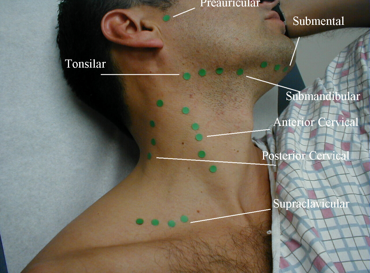
Intramammary lymph nodes IMLN are one of the most common benign findings at screening mammography. TB sarcoidosis lymphoma histoplasmosis and neoplasia are the common causes of mediastinal lymphadenopathy. Cervical lymph node levels IVI must be evaluated and documented bilaterally.
Idiopathic multicentric Castleman disease iMCD is a subtype of Castleman disease also known as giant lymph node hyperplasia lymphoid hamartoma or angiofollicular lymph node hyperplasia a group of lymphoproliferative disorders characterized by lymph node enlargement characteristic features on microscopic analysis of enlarged lymph node tissue and a range of. There were abnormal right axillary lymph nodes with increased eccentric hypoechoic cortical thickening measuring up to 5 mm above the normal value of 23 mm used at our institution Royal Free London NHS Trust Figure 3b.

Terms Definitions And Measurements To Describe Sonographic Features Of Lymph Nodes Consensus Opinion From The Vulvar International Tumor Analysis Vita Group Fischerova 2021 Ultrasound In Obstetrics Amp Gynecology Wiley Online Library
Lymph Nodes Swollen Lymph Nodes La Vascular
Cancers Free Full Text Oncologic Imaging Of The Lymphatic System Current Perspective With Multi Modality Imaging And New Horizon Html

Lymph Node Enlargement Radiology Reference Article Radiopaedia Org
Sonographic Appearance Of Abnormal Cervical Lymph Nodes In The Preoperative And Reoperative Empty Neck A Surgeon S Perspective Radiology Key

Normal Head And Neck Lymph Nodes In The Paediatric Population Clinical Radiology
Pin On Radiologia De Mama

Three Dimensional Sonography Of Axillary Lymph Nodes In Patients With Breast Cancer Koenigsberg 2016 Journal Of Ultrasound In Medicine Wiley Online Library

Abnormal Lymph Nodes Characteristics Concerning For Malignancy A Download Scientific Diagram

Lymph Nodes Radiology Key

Two Enlarged Inflammatory Reactive Perihepatic Lymph Nodes The Oval Download Scientific Diagram

Ultrasound Of The Right Axilla Shows An Enlarged Lymph Node With Download Scientific Diagram

Interobserver Variability Between Experienced Radiologists In Evaluating The Number Of Abnormal Lymph Nodes Seen On Preoperative Axillary Ultrasound Clinical Radiology
Uc San Diego S Practical Guide To Clinical Medicine

Cystic Lymph Nodes In The Lateral Neck As Indicators Of Metastatic Papillary Thyroid Cancer Endocrine Practice

Terms Definitions And Measurements To Describe Sonographic Features Of Lymph Nodes Consensus Opinion From The Vulvar International Tumor Analysis Vita Group Fischerova 2021 Ultrasound In Obstetrics Amp Gynecology Wiley Online Library

Ultrasonographic Differentiation Of Benign From Malignant Neck Lymphadenopathy In Thyroid Cancer Kuna 2006 Journal Of Ultrasound In Medicine Wiley Online Library

Sonography Of Neck Lymph Nodes Part Ii Abnormal Lymph Nodes Clinical Radiology

Metastatic Lymph Nodes At Gray Scale Examination In Patients With Download Scientific Diagram


Comments
Post a Comment